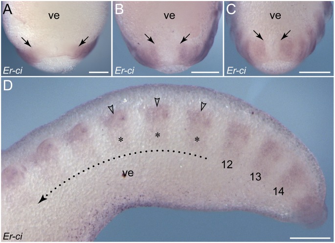
Figure 7. Details of cubitus interruptus expression in embryos of E. rowelli.
(A–C) Posterior ends of embryos at developmental stages II, III and IV in ventral view. Arrows indicate the posterior-most regions of expression, which subsequently move towards the proctodaeum. (D) Posterior end of a stage IV embryo in lateral view. Leg-bearing segments and corresponding limbs are numbered. Dotted line with an arrow indicates the direction of decreasing expression in the ventral ectoderm (asterisks) but increasing expression in the limb anlagen (arrowheads) towards the anterior end. Abbreviation: ve, ventral extra-embryonic tissue. Scale bars: 250 µm (A–D).

