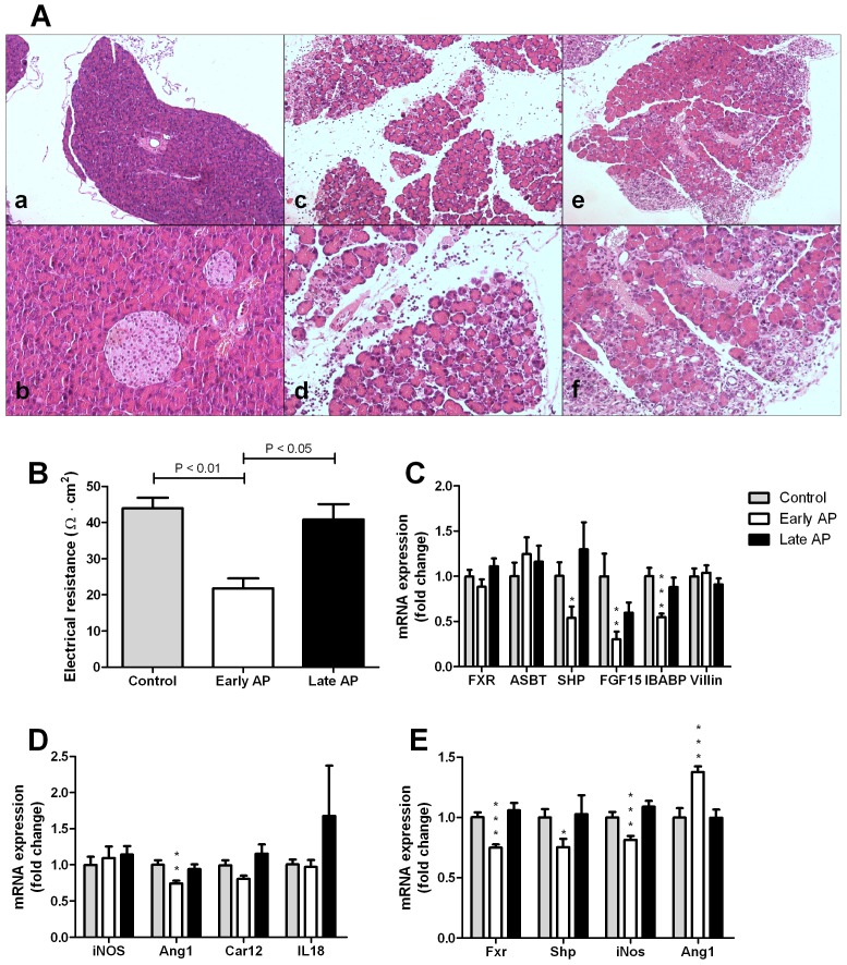
Figure 1.
A – Representative pancreatic histology of wild-type mice from the control group (a, b) and mice with early (c, d) and late (e, f) acute pancreatitis (H&E staining, 20x and 100x magnifications consecutively). Control mice have normal pancreatic morphology, whereas mice of the early pancreatitis group exhibit edema, influx of neutrophils and necrosis. Mice of the late pancreatitis group have no edema or necrosis, but show influx of lymphocytes and fibroblasts. B – Transepithelial electrical resistance of the ileum measured by Ussing chamber experiments. The resistance of the ileum was lower in the early pancreatitis group in comparison to both controls and the late pancreatitis group. C – Ileal mRNA expression of Fxr and FXR target-genes Asbt, Shp, Fgf15, and Ibabp, and Villin in wild-type mice of the control group, and the early and late pancreatitis groups. Expression of Fxr, Asbt and Villin did not differ between experimental groups. Expression of the other Fxr target genes was lower in early acute pancreatitis, but not in late pancreatitis. D – Ileal mRNA expression of genes implicated in intestinal barrier function, iNos, Ang1, Car12, IL18. Expression of Ang1 was lowered in the early pancreatitis group, the other genes remained similar in the different experimental conditions. E – Hepatic expression of Fxr, Shp, iNos and Ang1. Hepatic Ang1 was increased in early pancreatitis, whereas the expression of the other genes was lowered in the early acute pancreatitis group. Expression levels were normalized to cyclophilin expression. Bars indicate means and SEM, * p<0.05, ** p<0.01, *** p<0.001.

