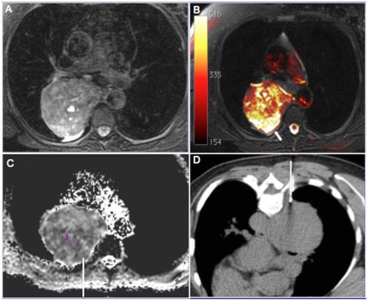
Figure 1.
A 62-year-old woman who presented with a heterogeneous mass in the right posterior mediastinum. The entire mass showed heterogeneous signal hyperintensity on T2-weighted magnetic resonance images (a) and fused T1- and diffusion-weighted images. (b) Note the remarkable signal hyperintensity in the posterior periphery of the lesion (arrow) compared with other areas, with definitive signs of restriction on the apparent diffusion coefficient (ADC) map. (c) The ADC value in the target area was 0.89×10⁻³ mm2/s. The needle was directed to this area during computed tomography–guided biopsy. (d) Histopathological analysis yielded a diagnosis of pleomorphic leiomyosarcoma.

