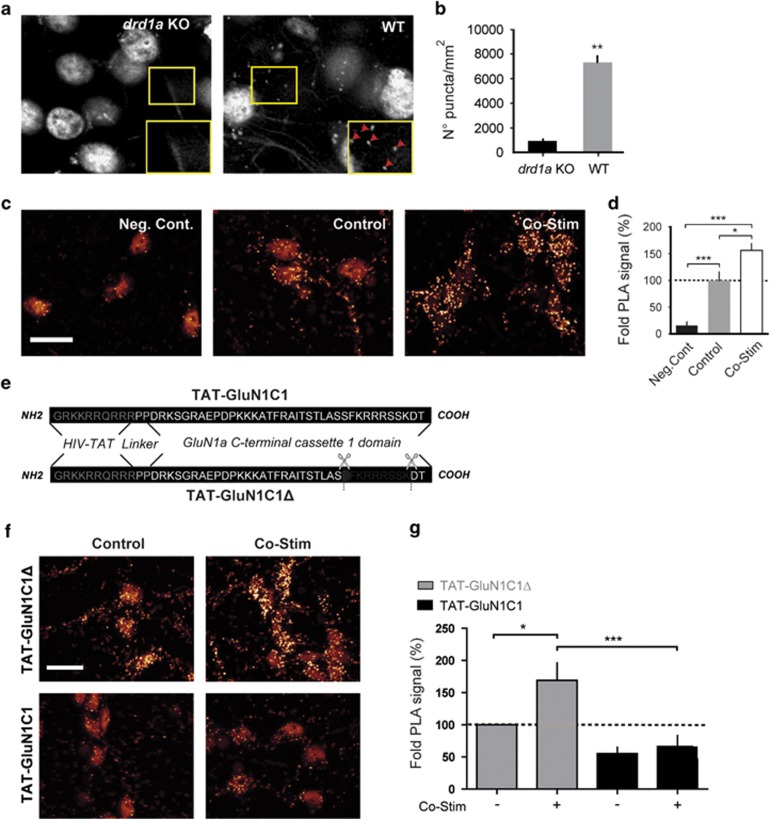
Figure 1.
Endogenous D1R/GluN1 complexes in medium spiny neurons (MSNs) are regulated by receptor co-stimulation and blocked by TAT-GluN1C1. (a) Confocal images and (b) quantification of D1R/GluN1 complexes detected by proximity ligation assay (PLA) in the nucleus accumben (NAc) of D1R-deficient (drd1a KO) or wild-type (WT) mice. **P<0.01; unpaired Student's t-test, n=4. (c) Images and (d) quantification of proximity ligation assay (PLA) signals from cultured MSN obtained when the dopamine D1 receptor (D1R) antibody is omitted as a negative control (Neg. Cont), or when the D1R and GluN1 antibodies are used after a 10-min incubation in the absence (Control) or presence of 0.3 μM glutamate and 3 μM SKF38393 (Co-stim). One-way analysis of variance (ANOVA), Newman–Keuls post hoc test, *P<0.05; ***P<0.001; N=4–5 independent experiments. (e) Amino-acid sequence of TAT-GluN1C1 and TAT-GluN1C1Δ peptides. (f) PLA images and (g) quantifications of D1R/GluN1 complexes from MSN pre-treated with TAT-GluN1C1Δ or TAT-GluN1C1 and co-stimulated or not one-way ANOVA, Newman–Keuls post hoc test, *P<0.05; ***P<0.001; n=5–6; unpaired Student's t-test. Scale bar, 10 μM.

