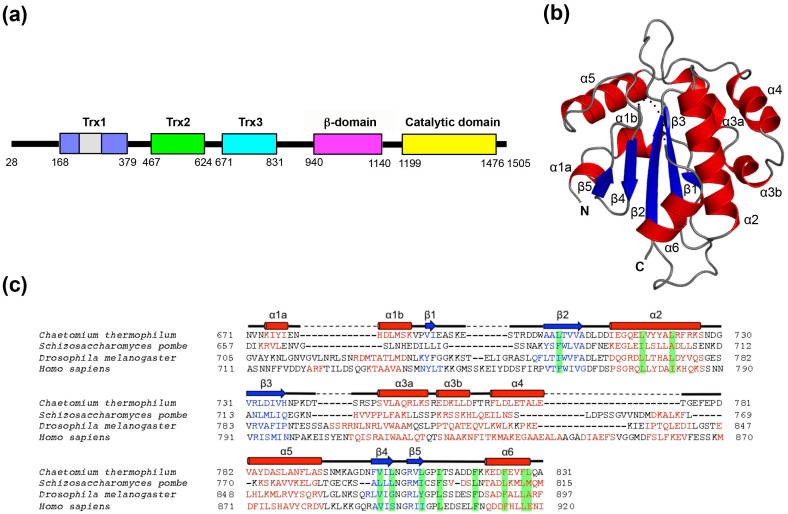
Figure 1.
Crystal structure of the Trx3 domain of UGGT (a) Domain structure of C. thermophilum UGGT. The Trx3 domain (residues Asn671–Ala831) was crystallized in this study. (b) Ribbon models of the Trx3 domain of C. thermophilum UGGT (Form 1). The secondary structures are highlighted (α-helix, red; β-sheet, blue) and the linker regions are shown in grey. The positions of the N- and C-termini are also indicated. Dotted line indicates disorder segment. (c) Structure-based sequence alignment of the Trx3 domains of UGGT among species (from fungi to human). The secondary structures of the Trx3 domain of C. thermophilum UGGT are indicated above the amino acid sequence. The secondary structure elements (α-helix and β-sheet) were predicted using the program PROMALS3D48 and are highlighted in red and blue, respectively. Residues involving the C-terminal α6 helix or detergent interactions are highlighted in green.

