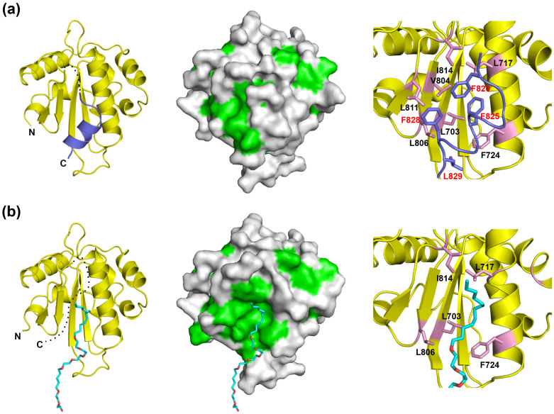
Figure 2. An extensive hydrophobic patch of the Trx3 domain is concealed by a flexible C-terminal helix.
The crystal structures of the Trx3 domain in Forms 1 and 2 are indicated in (a) and (b), respectively. The ribbon and surface models are shown in the left and centre. Dotted lines indicate disordered segments. In the surface model (centre), the hydrophobic residues are shown in green. Close-up views of the C-terminal helix or detergent-interacting regions are represented on the right. Residues involved in these interactions are highlighted in the pink stick model. In Form 1 (a), the C-terminal α6 helix is highlighted in slate. In Form 2 (b), the detergent ANAPOE C12E8 is shown as a stick model.

