
Figure 2.
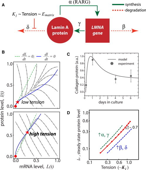
Feedback-based mechanobiological gene circuit model for lamin A exhibits polymer-physics scaling if cell tension suppresses protein turnover. (A) Nucleoskeletal Lamin-A protein regulates its own message (LMNA) and assembles in response to tension from matrix elasticity (Ematrix), which inhibits protein degradation. (B) Trajectories of lamin-A message and protein as the model converges from a range of initial conditions (arrowed lines) to where mRNA (dashed lines) and protein (solid lines) nullclines intersect to give a stable steady-state solution (star) appropriate to the tension (top, Kl = 0.5; bottom, Kl = 1.1). Null solution is unstable (hexagon). (C) Procollagen-1 expression in mesenchymal stem cells over seven days was tracked by immunolabeling and quantified. Stem cells adhere and spread on a substrate and start to synthesize collagen matrix, which stabilizes over time. The biphasic kinetic response can be recapitulated by the model that assumes tension-based inhibition of collagen protein degradation rate. (D) Setting the kinase/protease binding coefficient, Kl, to be proportional to (Tension)0.3 allows the model to generate steady-state scaling with tension that is consistent with tissue-level scaling of lamin A (4). Increasing the synthesis (α, γ) or degradation (β, δ) rates shifts steady-state lamin-A levels higher or lower, respectively. To see this figure in color, go online.