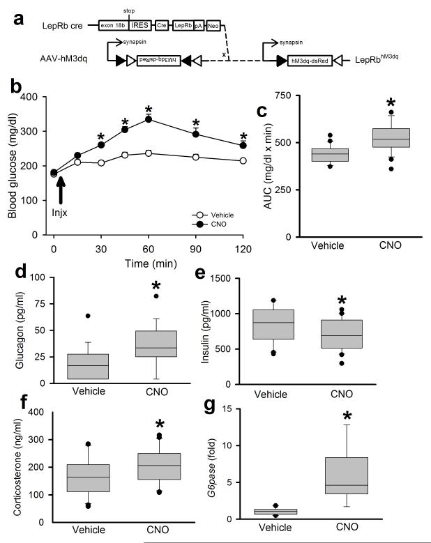
Figure 5. Activation of PBN LepRb neurons mimics the CRR to glucoprivation.
(a) Bilateral injection of the cre-inducible AAV-hM3Dq into the PBN of male Leprcre mice induces the expression of the activating DREADD hM3Dq in PBN LepRb neurons. Animals were injected with Vehicle or CNO (0.3 mg/kg, IP) and monitored for 120 minutes. (b) Blood glucose was monitored at the indicated times *p<0.001 (15min), <0.001 (30min), <0.001 (45min), <0.001 (60min), <0.001 (90min), 0.002 (120min) {F(6,30)=8.23}, N=16(veh) and 16(cno) animals. (c) Area under the curve (AUC) for the data graphed in (b); *p<0.001 {t(42)=−4.38} N=22 (veh) and 22 (cno) animals. Plasma glucagon (d) *p=0.002 {t(35)=−3.03} N=18(veh) and 19(cno) animals, insulin (e) *p=0.049 {t(40)=1.69} N=21(veh) and 21(cno) animals, corticosterone (f) 30 minutes after CNO administration *p=0.034 {t(40)=−1.87}, N=21(veh) and 21 (cno) animals. (g) hepatic G6Pase mRNA expression (fold over control) at the end of the experiment *p<0.001 {t(17)=−6.64}, N=10 (veh) and 9 (cno) animals. all data are plotted as mean +/− SEM. *p<0.05 The data in b were analyzed by two way repeated measures ANOVA with Fisher LSD post hoc test. The data in c were analyzed by two tailed t-test; d-g were analyzed by one tailed t-test, due to our directional hypothesis. Data in b is plotted as mean +/− SEM and as Q1, Q2, and Q3 with whiskers at the 10th and 90th percentiles in c-g.

