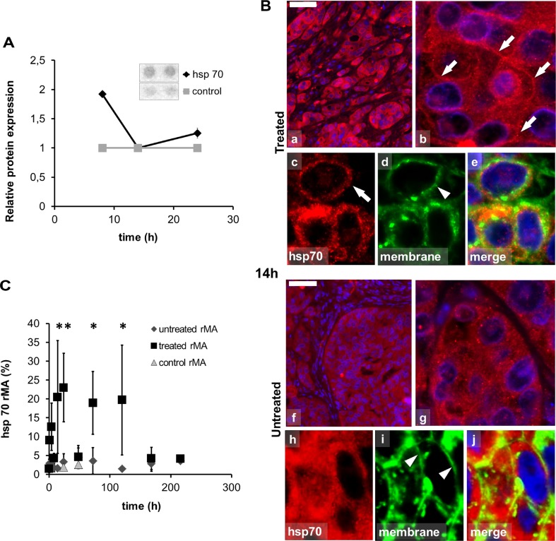
Fig. 2.
Abundance and localization of heat shock protein 70 (Hsp70) in colorectal cancer xenografts measured with apoptosis protein array and immunofluorescence (Alexa 564, red). a Pooled tissue samples reveal elevated Hsp70 levels in apoptosis protein array at 8 h with no change at 14 and 24 h due to inhomogeneity of the tissue at these time points. b Hsp70 immunofluorescence (Alexa 564, red), in 14-h post-mEHT treated (a–e) and untreated (f–j) tumor cells also immunoreacted with wheat germ agglutinin (Alexa 488, green) and DAPI to stain nuclei. In 14 h post-mEHT-treated tumor cells Hsp70 immunofluorescence is predominately in cell membranes (arrows) as indicated by the wheat germ agglutinin reactivity (arrowheads). In the untreated tumor cells Hsp70 immunofluorescence is predominately cytoplasmic. Scale bar = 80 μm in a and f, 10 μm in b and g, and 5 μm in c, d, e, h, i, and j. c Semi-quantitative image analysis of immunofluorescence confirms significant elevation of Hsp70 levels both between 14–24 h and 72–120 h post-treatment (*p < 0.05) with a decline at 48 h (rMA relative mask area)

