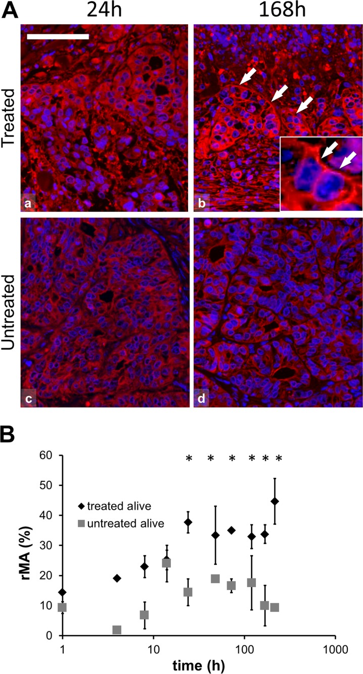
Fig. 3.
Hsp90 immunofluorescence (Alexa 564, red) and semi-quantitative analysis of heat shock protein 90 (Hsp90) in post-mEHT treated and untreated colorectal cancer xenografts. a Hsp90 immunofluorescence is predominately cytoplasmic at 24 h (a) and associated with cell membranes at 168 h post-mEHT (b; arrows), and as shown in the inset. Hsp90 immunofluorescence is less apparent at 24 h (c) and at 168 h (d) in untreated tumor cells. Cell nuclei are stained blue (DAPI). Scale bar = 60 μm in all and 20 μm in the inset. b Graph showing significant increase of Hsp90 protein between 24 and 216 h in the treated compared to the untreated tumor cells in the morphologically intact tumor areas (*p < 0.05) (rMA relative mask area)

