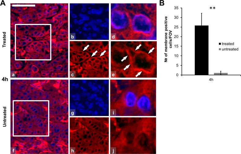
Fig. 4.
Calreticulin immunofluorescence (Alexa 564, red) and semi-quantitative analysis of calreticulin in post-mEHT treated and untreated colorectal cancer xenografts. a Calreticulin immunofluorescence is localized to the cell membranes 4 h post-mEHT (arrows; a–e) before any morphological or molecular sign of programmed cell death. Calreticulin immunofluorescence is diffuse and cytoplasmic in untreated tumor cells (f–j). Cells nuclei are stained blue (DAPI). Scale bar = 60 μm in a, b, c, f, g, and h and 10 μm in d, e, i, and j. b Graph showing the mean number of cytoplasmic membrane positive cells counted at ×100 magnification in 10 fields of views (FOV) of five parallel samples. Elevation of calreticulin cell membrane immunofluorescence is highly significant (**p < 0.01) in 4 h post-mEHT treated compared to untreated tumor cells (FOV field of view)

