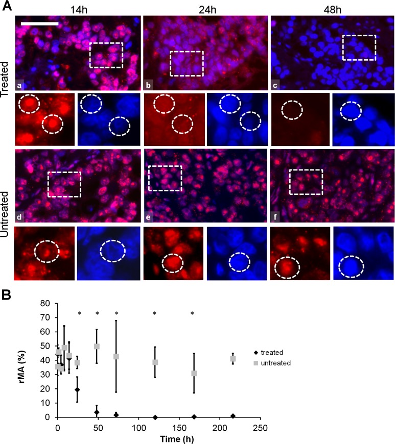
Fig. 5.
HMGB1 immunofluorescence (Alexa 564, red) and semi-quantitative analysis of HMGB1 post-mEHT treated and untreated colorectal cancer xenografts. a HMGB1 immunofluorescence shows normal nuclear localization up to 14 h post-mEHT treated (a) and untreated (d) tumor cells. HMGB1 immunofluorescence is diffuse and diminished in 24 h and not evident in 48 h post-mEHT treated (b and c, respectively) tumor cells but not changed in 24 and 48 h untreated (e and f, respectively) tumor cells. Boxes show the location of the high-magnification insets. Circles in the insets show the location of nuclei. Scale bar = 60 μm in a, b, c, d, e and f, and 25 μm in all insets. b Semi-quantitative analysis highlights significant reduction of HMGB1 immunofluorescence between 48 and 216 h post-mEHT treated compared to untreated tumor cells (*p < 0.05) (rMA relative mask area)

