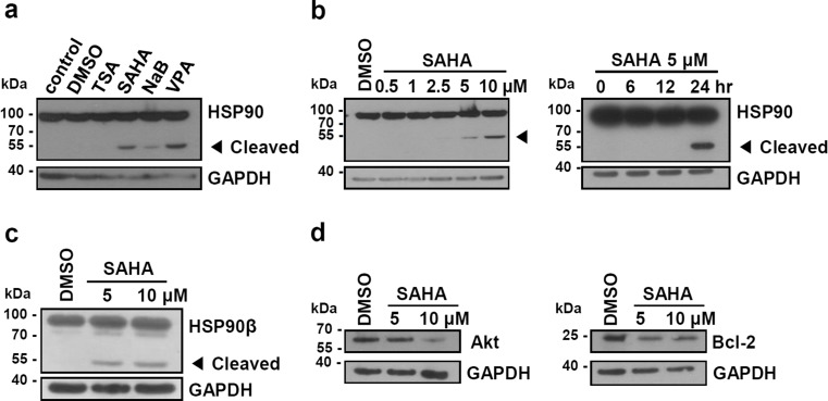
Fig. 1.
HDAC inhibitors induce cleavage of HSP90. a K562 cells were treated with HDAC inhibitors (250 nM TSA, 5 μM SAHA, 4 mM NaB, 4 mM VPA) for 24 h. b K562 cells were treated with the indicated doses of SAHA for 24 h and with 5 μM SAHA during the indicated periods. c, d K562 cells were treated with the indicated dose of SAHA for 24 h. The cell lysates were subjected to Western blot analysis using anti-HSP90, anti-HSP90β, anti-Bcl-2, anti-Akt, and anti-GAPDH antibodies. Amounts of GAPDH protein are shown as a loading control. Cleaved HSP90 fragment is indicated by an arrowhead

