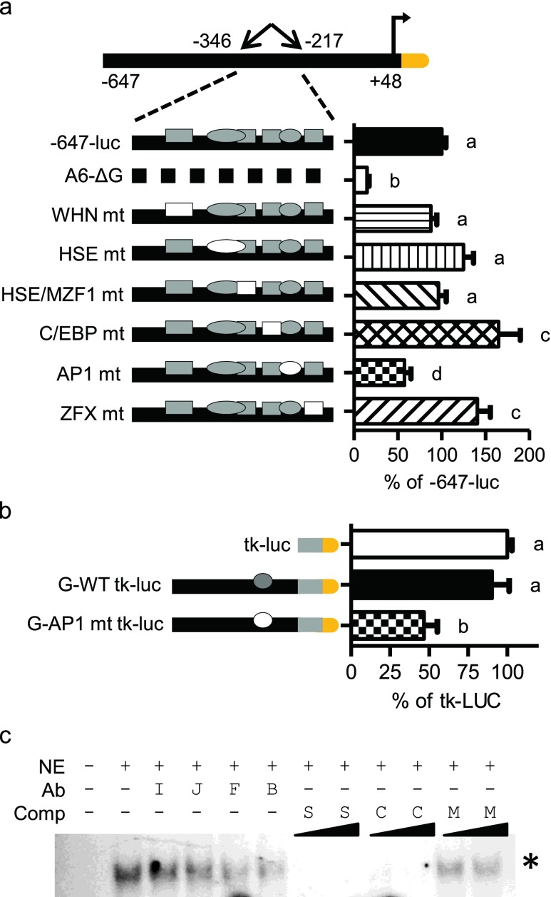
Fig. 5.
Characterization of the −244 bp AP1 site. a Site-specific mutations within the −647-luc construct. Filled shapes indicate wild-type elements. Specific site mutants are shown as empty shapes. Graph shown as percent of −647-luc. b AP1-specific mutant within the fragment G-tk-luc construct. Graph shown as percent of tk-luc. Statistically significant values are indicated by different letters using a p value <0.05 as determined using ANOVA with Newman Keuls post hoc. Bars are mean + SEM from three experimental repeats, each bar from triplicate cultures. c EMSA of AP1. Antibodies include nonspecific IgG (I), anti-cJun (J), anti-cFos (F), or both anti-cJun and anti-cFos (B). Nonradiolabeled competitor oligomers include self HSPA6 AP1 site (S), consensus oligomer (C), or mutated AP1 (M) at 5- or 50-fold excess. Asterisk denotes disrupted binding species

