
Fig. 1.
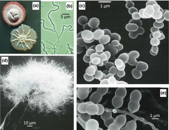
Morphological diversity of actinobacteria. (a) Colonies of Streptomyces coelicolor A3(2) wild type (upper) and bldA mutant (lower). (b) Phase contrast image of sporulating mycelium of S. coelicolor. (c) Micrococcus luteus. (d) Actinosynemma mirum. (e) Microbispora rosea. Images [c–e] are scanning electron micrographs taken from Miyadoh (1997), with permission.