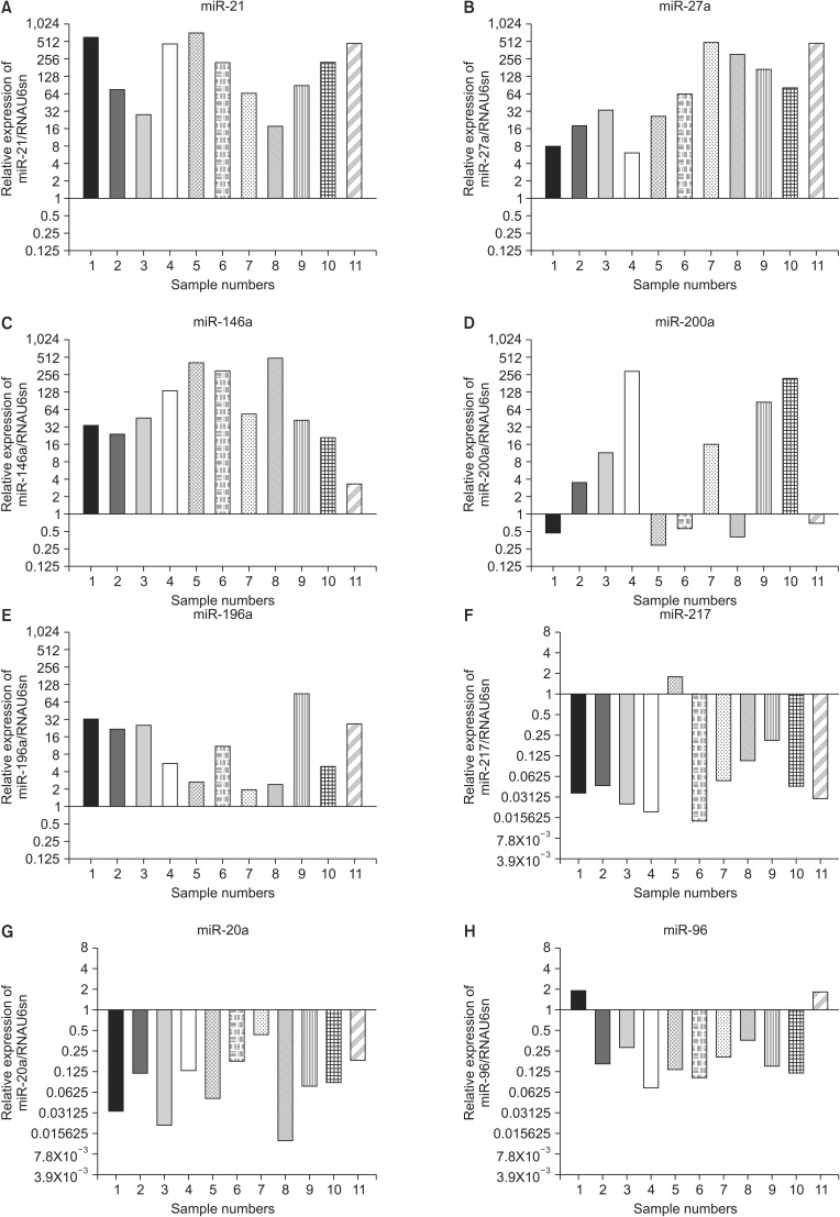
Fig. 2.
Comparative expression analysis of miR-21 (A), miR-27a (B), miR-146a (C), miR-200a (D), miR-196a (E), miR-217 (F), miR-20a (G), and miR-96 (H) levels in 11 fine-needle aspiration specimens from cancer relative to matched normal control tissue, as estimated by quantitative RT-PCR. The expression levels in control paraneoplastic normal pancreatic tissues were set to 1.0 in terms of log2 values.

