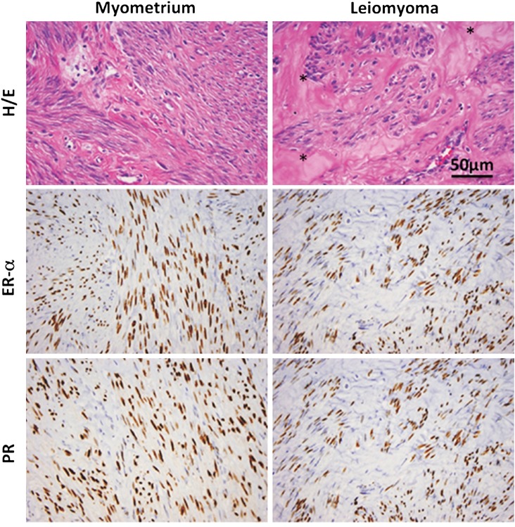
Figure 1.
Typical histology of human leiomyoma. Representative slides demonstrating the typical hematoxylin/eosin (H/E) slide and immunohistochemistry stains for estrogen receptor-α (ER-α) and PR in myometrium and leiomyoma; *denotes extracellular matrix (ECM) in the H/E-stained leiomyoma section. Note the expanded ECM leiomyoma tissue and increased nuclear size in leiomyoma smooth muscle cells. Immunoreactive ER and PR (brown stain) are localized to the nuclei of myometrial or leiomyoma smooth muscle cells. Images courtesy of Dr Jian-Jun Wei, Department of Pathology, Northwestern Memorial Hospital, Chicago, IL, USA. Scale bar represents 50 μm.

