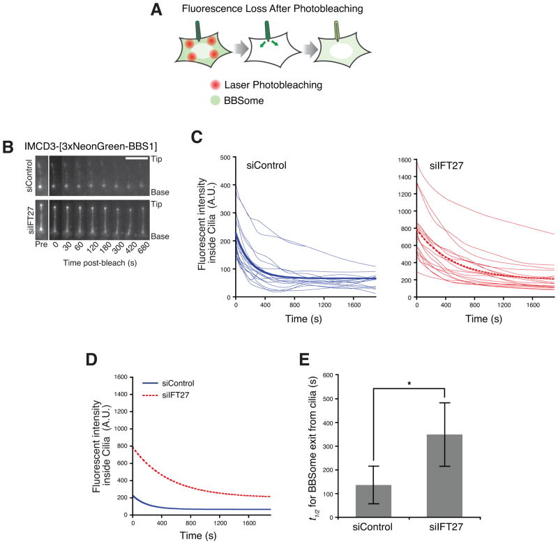
Figure 5. IFT27 is required for rapid exit of BBSome from cilia.
(A) Schematic of Fluorescence Loss After Photobleaching (FLAP) assay. NG3-BBS1 was photobleached in the cytoplasm by intense illuminations with a 488 nm laser. The bleached areas of the cell are distant from the cilium to ensure that ciliary NG3-BBS1 is not bleached by the illuminations. The subsequent loss of NG3-BBS1 fluorescence from cilia was monitored by live imaging.
(B) Time series montage representing the dynamic loss of ciliary NG3-BBS1 fluorescence in FLAP assay. Ciliary tip and base are marked. Scale bar, 5 μm.
(C) Decay of ciliary NG3-BBS1 fluorescence signal in FLAP assays for control siRNA- and IFT27 siRNA-treated cells. The fluorescence decay was measured for individual cilia, and plotted as a smoothed line for siControl (left, blue lines) and siIFT27 (right, red lines) treated cells. Photobleaching was negligible (<2%, data not shown). Each experiment was individually fit to a single exponential, and a simulation describing the average of these fits is shown as a bold line. Data were collected from five independent experiments (number of cilia analyzed n=17 for siControl and n=18 for siIFT27).
(D and E) The exit of BBSome from cilia is slower in the absence of IFT27. (D) Replotting of the simulations describing the average fits from panel (B) for siControl (blue solid line) and siIFT27 (red dotted line) treated cells. (E) Average half-lives (t1/2) for ciliary exit of NG3-BBSome. For siControl, t1/2 =136s +/− 20s, and for siIFT27, t1/2 =349s +/− 20s. The asterisk indicates a highly significant difference in exit rates (unpaired t-test, p < 5 × 10⁻⁶). Error bars represent +/− 1 SD.

