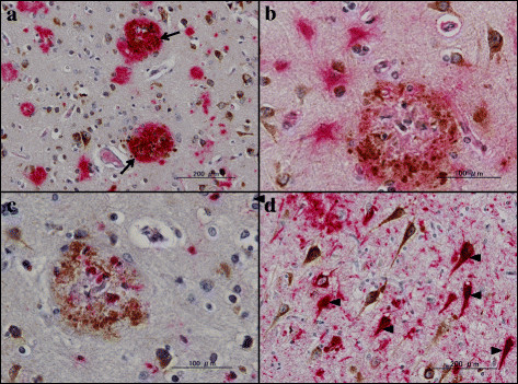
Figure 5.

PLD3 immunoreactivity in Alzheimer’s disease brains (II). PLD3 immunoreactivity was studied in Alzheimer’s disease brains by double-labeling immunohistochemistry. (a) Frontal cortex, PLD3 (brown), amyloid beta (Aβ; red), colocalization of PLD3 and Aβ (arrows). (b) Frontal cortex, PLD3 (brown), GFAP (red), reactive astrocytes do not express PLD3. (c) Frontal cortex, PLD3 (brown), CD68 (red), activated microglia do not express PLD3. (d) Hippocampal CA1 region, PLD3 (brown), AT8-tau (red), colocalization of neurofibrillary tangles and PLD3 (arrowheads). PLD, phospholipase D.
