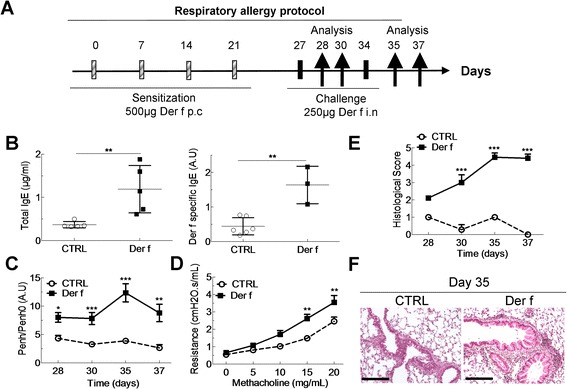
Figure 2.

Mouse model of Der f-induced asthma defined by hyperresponsiveness and pulmonary infiltrate. (A) Balb/c mice sensitized to Der f by percutaneous (p.c) application on days 0, 7, 14 and 21 and then challenged intranasally (i.n) with Der f on day 27 and 34. Mice were sacrificed after one or three days after the first (day 28 and 30) or second challenge (day 35 and 37). On day 35, blood was removed and serum collected to measure (B) total IgE, Der f-specific IgE, by ELISA in control (white circles) and Der f-allergic mice (black squares). (C) Measurement of airway hyperresponsiveness (AHR) after one and three days after the first (day 27) and second challenge (day 34) in control (CTRL, white circle dotted line) and in allergic (Der f, black square plain line) mice. AHR is displayed by Penh/Penh0 (pause enhanced ratio to basal pause enhanced). (D) Airway resistances to increasing doses of methacholin on day 37 in CTRL and Der-f sensitized mice. (E) Inflammatory score in lungs one and three days after the first (day 27) and second challenge (day 34) in CTRL and Der f mice. (F) Representative hematoxylin-eosin staining of a lung section in control (CTRL) and allergic mice (Der f) on day 35. Scale bars represent 100 μm. Data are represented as mean ± SEM (n = at least 5 mice/group). *p < 0.05, **p < 0.01, ***p < 0.001.
