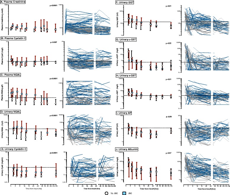
Figure 2.

Temporal profiles of acute kidney injury (AKI) biomarker concentrations in patients with and without AKI. For each biomarker the graph on the right has individual profiles of AKI patients (blue lines) and non-AKI patients (grey lines). The graph on the left represents median values of AKI patients (blue squares), and non-AKI patients (white circles). Vertical bars cover the interquartile range. The dashed horizontal lines are pre-specified cut points of biomarkers. P-values are for comparison of biomarker concentrations between those with and without AKI were performed using repeated-measures analysis of variance after log-transformation. (A) Plasma creatinine; (B) plasma cystatin C; (C) plasma neutrophil gelatinase-associated lipocalin (NGAL); (D) urinary NGAL; (E) urinary cystatin C; (F) urinary gamma-glutamyltranspeptidase (GGT); (G) urinary α-glutathione-S-transferase (GST); (H) urinary π-GST; (I) urinary alkaline phosphatase (AP); (J) urinary albumin.
