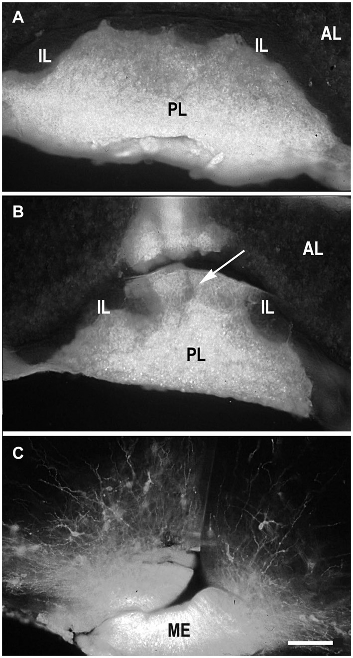
Figure 1.

DiI labeling of the posterior lobe of the pituitary and median eminence. DiI distribution in the pituitary after insertion into the posterior pituitary lobe (PL) without damage of the boundary between the PL and intermediate (IL) pituitary lobes (A). DiI insertion with labeling in the PL and part (arrow) of IL (B). DiI insertion in the median eminence (ME) with few labeled neurons in the arcuate nucleus (C). AL – anterior pituitary lobe. Scale bar = 100 μm.
