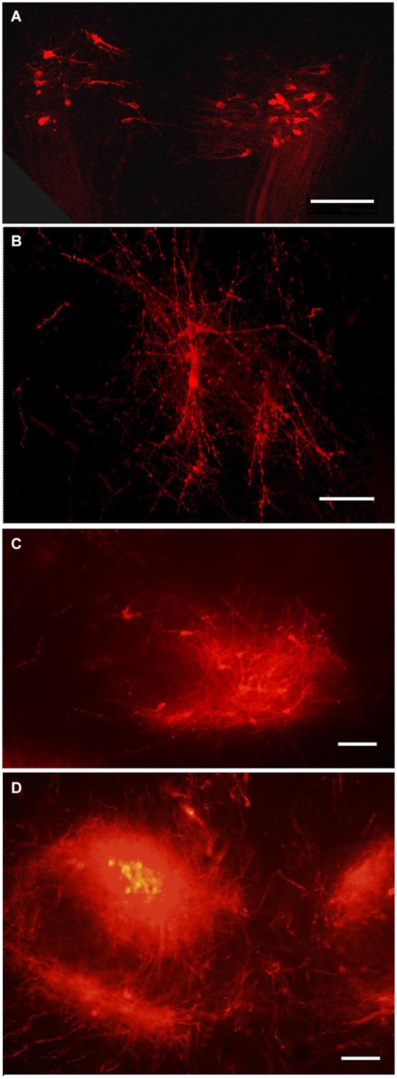
Figure 5.

Tegmental neurons innervating MBO. DiI labeled neurons in the tegmentum following DiI insertion into the MBO on different prenatal stages: (A) E14, bilateral groups of neurons on a coronal section; (B) E19, confocal image of the same neurons, in sagittal section; (C) P1, the ventral tegmental nucleus on sagittal section; (D) P10, ventral tegmental nuclei on coronal section. Scale bars: (A,B) = 80 μm, (C,D) = 100 μm.
