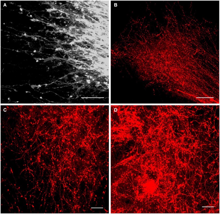
Figure 6.
Innervation of the anterior thalamic complex by the MBO. High-resolution confocal images of the fine details of the growing mammillothalamic tract and innervation of the anterior thalamic nuclei following DiI insertion in the mammillary body. (A) E19.5, axonal growth cones on the top of MTh. (B) P2, rostral end of the MTh growing into the anteromedial thalamic nucleus and forms terminal arborizations. (C) P4 and (D) P10, terminal network of the mammillary body axons in the anteromedial and anteroventral thalamic nuclei. Scale bars: (A,C,D) = 20 μm, (B) = 80 μm.

