
Fig 6.
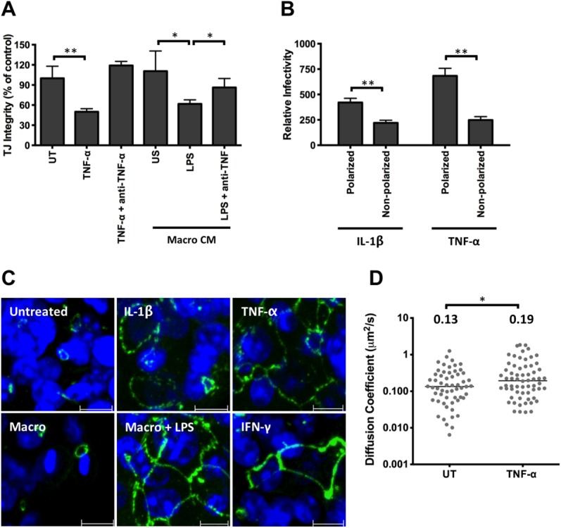
Effect of TNF-α on HepG2 permeability, occludin localization, and CD81 lateral diffusion. (A) Polarized HepG2.CD81 were treated with cytokines or CM from unstimulated (US) or LPS-stimulated macrophages, with or without anti-TNF-α (100 μg/mL) and tight junction integrity quantified. (B) Effect of IL-1β or TNF-α on HCVpp infection of polarized or nonpolarized HepG2.CD81. Data are expressed relative to untreated (100%) polarized or nonpolarized control. (C) Occludin localization in HepG2.CD81 cells following cytokine or CM treatment, where scale bar = 10 μm. (D) AcGFP-CD81 diffusion speed in untreated (UT) or TNF-α treated polarized HepG2 cells, where each symbol represents an independent bleach spot. The median values are shown. **P < 0.01, *P < 0.05.