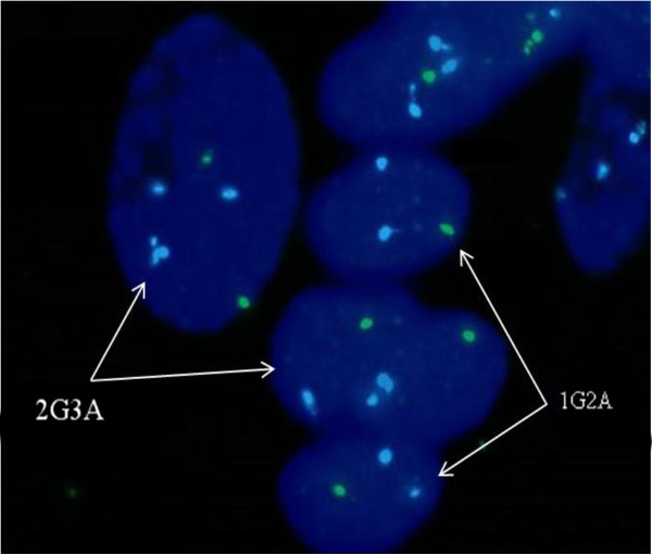
Figure 4.

FISH analysis results of placental tissue showed the combination status of 45, X and trisomy 18 mosaicism, with DNA of chromosome 18 marked as aqua (A) and chromosome X as green (G). The cells with karyotype 47, XX, +18 were indicated as 2G3A, while the 45, X cells were indicated as 1G2A. Two additional chromatin segments attached to a chromosome 18 were observed in a cell (2G3A) on the left, suggesting partial duplications in addition to trisomy 18.
