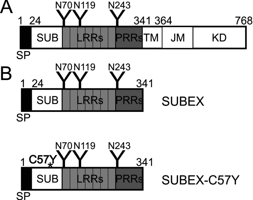
Figure 1. Domain structure of SUB and SUBEX variants.
(A) Schematic representation of the full-length SUB protein. Different protein domains are indicated [22]. (B) Schematic representation of the truncated proteins SUBEX and SUBEX-C57Y, comprising only the extracellular domain of SUB. The C57Y mutation is indicated by an asterisk. N-glycosylation sites are represented by ‘Y’ and their amino acid positions are shown. JM, juxtamembrane domain; KD, kinase domain; LRR, leucine-rich repeat; PRR, proline-rich repeat; SP, signal peptide; SUB, SUB-domain; TM, transmembrane domain.

