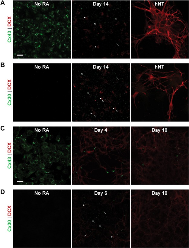
Figure 2.

Spatial expression pattern of Cx43 and Cx30 protein with DCX as a marker of differentiating neurons in the NT2/D1 and P19 EC cell models. Scale bar = 20 µm. (A) Representative images of ICC labelling of Cx43 (green) and DCX (red) in undifferentiated NT2/D1 cells, cells at day 14 of RA treatment and mature hNT neuronal cultures. Intracellular and punctate labelling of Cx43 was observed in undifferentiated cells, however, immunolabelling for Cx43 was much less prevalent at day 14 (arrows) and appeared to largely be on cells without strong DCX staining rather than DCX-positive cells (arrowheads). In mature hNT neurons, Cx43 was not observed, while strong DCX was present in both cell bodies and neurites. (B) Representative images of ICC labelling of Cx30 (green) and DCX (red) in undifferentiated NT2/D1 cells, cells at day 14 of RA treatment and mature hNT neuronal cultures. Cx30 was not observed in undifferentiated NT2/D1 cells, however puncta became widespread peaking at day 14 of RA treatment, before decreasing again. Cx30 was not observed in cultures of hNT neurons. Cx30 puncta (arrows) were visible on DCX-positive cells (arrowheads) and on cells absent of DCX staining at day 14, with no obvious preferential localisation. (C) Representative images of ICC labelling of Cx43 (green) and DCX (red) in undifferentiated P19 EC cells, cells at day 4 post-RA treatment and day 10 post-RA treatment. Similar to NT2/D1 cells, Cx43 was observed as both intracellular and punctate labelling. Cx43 was initially ubiquitously expressed, however, both punctate and intracellular labelling decreased after RA treatment by day 4 post-RA treatment and remained low at day 10 post-RA treatment. (D) Representative images of ICC labelling of Cx30 (green) and DCX (red) in undifferentiated P19 EC cells, cells at day 6 post-RA treatment and day 10 post-RA treatment. Cx30 was not observed in undifferentiated P19 cells. At the 6-day timepoint post-RA treatment, Cx30 puncta were observed (arrows) at the centre of large clusters of cells with DCX-positive neurites (arrowheads) extending from them at their periphery. This peak in expression was transient as Cx30 decreased between day 6 and day 10 post-RA treatment.
