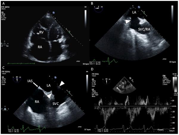
Figure 4.

Representative images from transthoracic echocardiography (TTE) and transesophageal echocardiography (TEE). A, TTE demonstrates marked right atrial (RA) and right ventricular (RV) dilatation. B, TEE image at a 5° omniplane angle and above the level of the aortic valve demonstrates an abnormal interatrial communication from the superior vena cava (SVC)–RA to the left atrium (LA; arrow). C, TEE image in a plane orthogonal to B, at a 93° omniplane angle, demonstrates a modified bicaval view showing structures superior to the interatrial septum (IAS). There is a communication from the LA to the SVC that occurs as a result of the absence of tissue separating the SVC from the LA (arrows). D, Pulsed-wave Doppler through the interatrial communication shows bidirectional but predominantly left-to-right flows.
