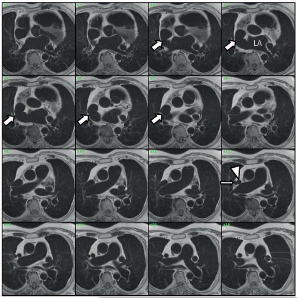
Figure 5.
Anomalous right superior pulmonary vein (RSPV) and superior sinus venosus defect are identified by cardiac magnetic resonance imaging (cMR). Serial axial cMR slices are shown, with the first image at the level of the aortic valve and sequential images obtained by moving superiorly. These images demonstrate both the anastomosis of the superior vena cava (SVC; arrowhead) and RSPV (thin arrow) and the presence of an abnormal communication between the left atrium (LA) and the SVC–right atrial junction (arrows). The morphology and geometry of the interatrial communication mirror the anatomy shown by transesophageal echocardiography. In addition, the pulmonary artery is dilated, and the persistent left SVC is visible.

