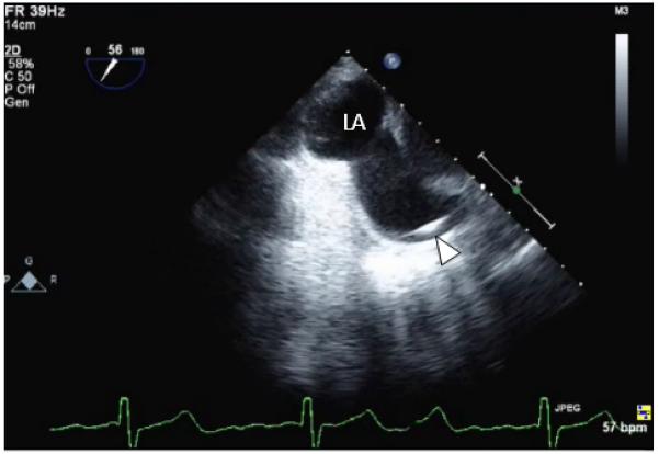
Figure 6.

Intraoperative transesophageal echocardiography (TEE) image before patch repair. A Swan-Ganz catheter is visible within the superior vena cava (SVC; arrowhead), and this TEE image at a 56° omniplane angle shows that the SVC communicates with the left atrium.
