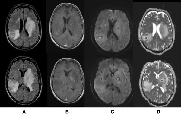
Figure 2.

Brain magnetic resonance imaging (MRI) on day 2. Showed multiple rounded expansive lesions in basal ganglia region predominantly on the left side and in subcortical right parietal region with prominent oedema, resulting in modelling of lateral ventricles, slight deviation of middle line to the right side and reduction of sulci dimensions (A: T2 flair). These lesions present a post gadolinium multilayered ring enhancement pattern (B: T1 post gadolinium) and central core with restrition of water molecules diffusibility (C: DWI, D: ADC map).
