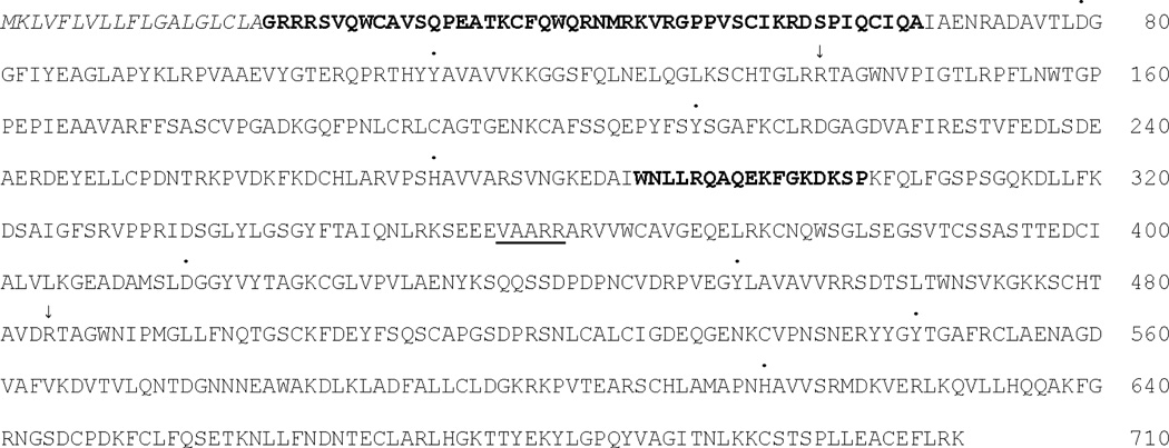
Figure 1.
Primary structure of human lactoferrin (NP_002334). The signal peptide is italicized; the two positively charged regions of the N-lobe are in bold face; the linker between the N-lobe and C-lobe is underlined. Dots above residues indicate iron-binding residues; arrows above residues indicate carbonate-binding residues.

