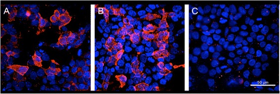
Figure 2.

Cell based assays for demonstration of CASPR2 antibodies (Euroimmun, Lübeck, Germany). (A) Serum of patient diluted 1:15 incubated with HEK cells transfected with CASPR2; the antibodies are visualized by a Alexa 594 anti-human-IgG antibody; mild counterstaining with Hoechst 33342. (B) Serum of a patient with classical limbic encephalitis and CASPR2 antibodies, technical details as in A. (C) Serum of patient incubated with control cells not expressing CASPR2 (negative result), technical details as in A. These images demonstrate that the patient’s serum does not bind non-specifically to the CASPR2 expressing HEK cells in A.
