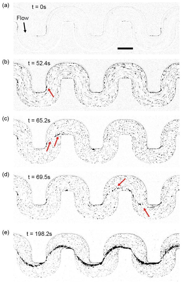
Figure 2.

Initiation of the S. aureus biofilm streamer and its rapid expansion. The displayed images are representative snapshots from a movie, acquired at 30 fps using bright field illumination. The images were taken at a focal plane that is perpendicular to gravity and in the middle of the channel, which is 100 μm from the bottom of the channel. (a) Initial image of the channel, which is coated with human blood plasma proteins. (b) A thin streamer originates from the corner on which surface-attached biomass has accumulated. (c) A streamer has bridged the gap between adjacent corners. At this stage, the streamer is flexible and vibrates in the flow. (d) Occasionally streamers become dislodged and they can reattach elsewhere downstream. (e) Stable streamers have formed on all corners and accumulated additional biomass, making them less flexible. The suspension we flowed through the channel was at a cell density that corresponds to OD600=1.2. The scale bar represents 200 μm.
