
Figure 3. Comparison of two different methods for measuring the opening/closing of the O-helix on DNA Polymerase I.
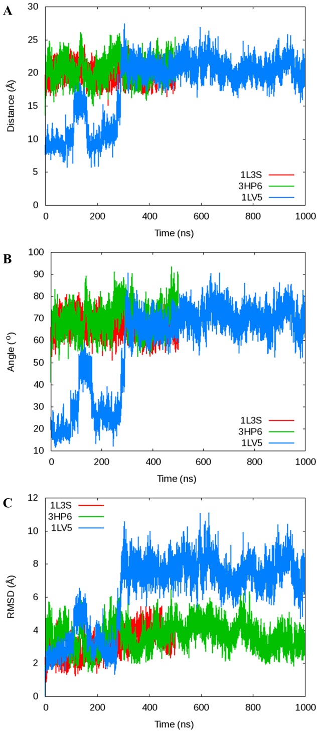
A) The α-C distance between Arg629 and Pro699 shown in this manuscript compared to B) the angle between the α-C of Arg629, Gly711, and Asn700 used by Golosov et al. to determine the conformation of the O-helix and C) a plot of the RMSD of the fingers domain as a function of time in reference to the original crystal structure used to start each simulation. The distance, angle, and RMSD measurements are directly comparable, validating our use of the Arg629-Pro699 distance.