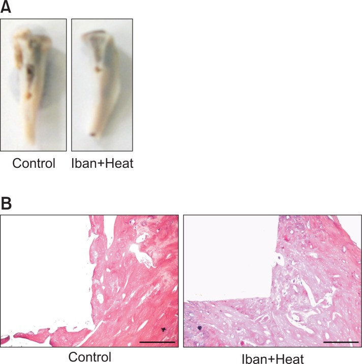
Fig. 1.
Photographs of bone after removing an implant. The bone was fixed according to the shape of the implant (A). Histological appearance of bone after staining with hematoxylin and eosin (B). The picture depicts the ongoing fixation of the bone to the implant. Photographs captured at 50X, and the scale bar is 50 μM.

