
Figure 5.
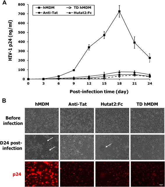
Reducing of HIV-1 replication by lentivirus-mediated expression of Hutat2:Fc in primary hMDM. (A) Kinetics of HIV-1Ba−L replications (HIV-1 p24 levels). The data showed a significant reduction of HIV-1 replication in both the TD-hMDM and Hutat2:Fc culture groups as compared to hMDM (P <0.01), but no statistical difference among TD-hMDM, Hutat2:Fc, and Anti-Tat groups (P >0.05). (B) Lentiviral vectors HR-Hutat2 transduction suppresses HIV-1 cytopathicity and the expression of p24 in hMDM cultures. Normal hMDM and HR-Hutat2 transduced hMDM were exposed to HIV-1Ba-L, and examined before and on day 24 post-viral infection using a 10× objective. It can be readily appreciated that either HR-Hutat2 transduction or Hutat2:Fc strongly suppressed HIV-1-mediated cytopathic effects, resulting in a reduction in the number of giant cells in the culture. In addition, HIV-1 p24 immunofluorescent staining showed that HR-Hutat2 transduction and Hutat2:Fc reduced the expression of HIV-1 p24 intracellularly. Images were acquired as described in Figure 1. hMDM, Normal hMDM; TD-hMDM, HR-Hutat2 transduced hMDM; Anti-Tat, Non-transduced hMDM treated with anti-HIV-1 Tat antibody; Hutat2,Fc, Normal hMDM treated with conditioned medium from HR-Hutat2 transduced hMDM; D24 post-infection, Day 24 post-HIV-1-infection; p24, HIV-1 p24 immunofluorescent staining; White arrow, HIV-1-induced cytopathic effect. The blood of three donors was used in this assay. Results represent mean values from triple independent experiments and error bars denote the s.e.m. Scale bar = 100 μm.