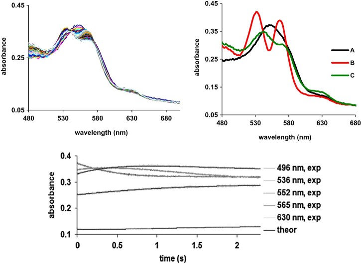
Fig. 6.
Upper panel: left UV-vis spectra of deoxyhemoglobin treated with nitrite, recorded for up to 2 seconds after mixing. Conditions: deoxyhemoglobin was obtained by purging with argon, and then adding a glucose oxidaze/glucose/catalase mixture as indicated in Experimental, buffer PBS, pH = 7.4, [Hb] = 18 µM, [nitrite] = 925 mM. Right: involved species as resulted from the fitted spectra, employing the A → B, B → C model. Lower panel: left: time evolution of the three fitted species, A, B and C; right: overlay of kinetic data and of fitted trace at some representative wavelength.

