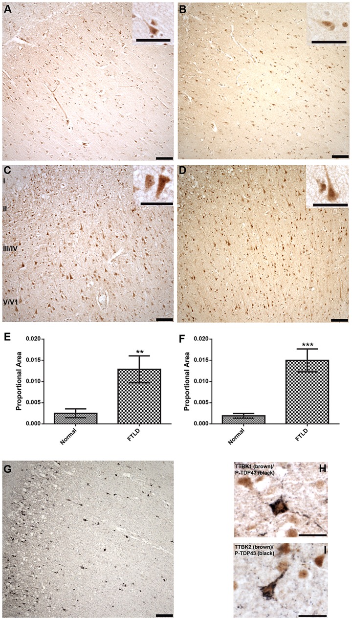
Figure 4. Upregulated Tau tubulin kinases are also co-expressed with phospho-TDP-43 pathology.
Representative photomicrographs depicting TTBK1 (A, C) and TTBK2 (B, D) immunoreactivity in cortical neurons in normal (A, B) and FTLD-TDP Type B (C, D) cases. The cellular distribution is both cytoplasmic and nuclear (insets), and immunoreactivity appears to be more widespread in FTLD cases relative to normal controls. Cortical layers I-VI are indicated (C). Quantification of immunostaining demonstrated a statistically significant increase in both TTBK1 (E) and TTBK2 (F) in FTLD cases compared to normal controls (**P = 0.003; ***P<0.0001). The distribution of phospho-TDP-43 immunoreactivity in the cortex of an FTLD case (G) overlaps with TTBK1 (C) and TTBK2 (D). Double label immunohistochemical experiments suggest co-localization of phospho-TDP-43 with TTBK1 (H) and TTBK2 (I) in an FTLD case. Scale bars: 100 µm A–D,G; 50 µm insets A–D; 25 µm H,I. See S5 Figure for controls for antibody specificity.

