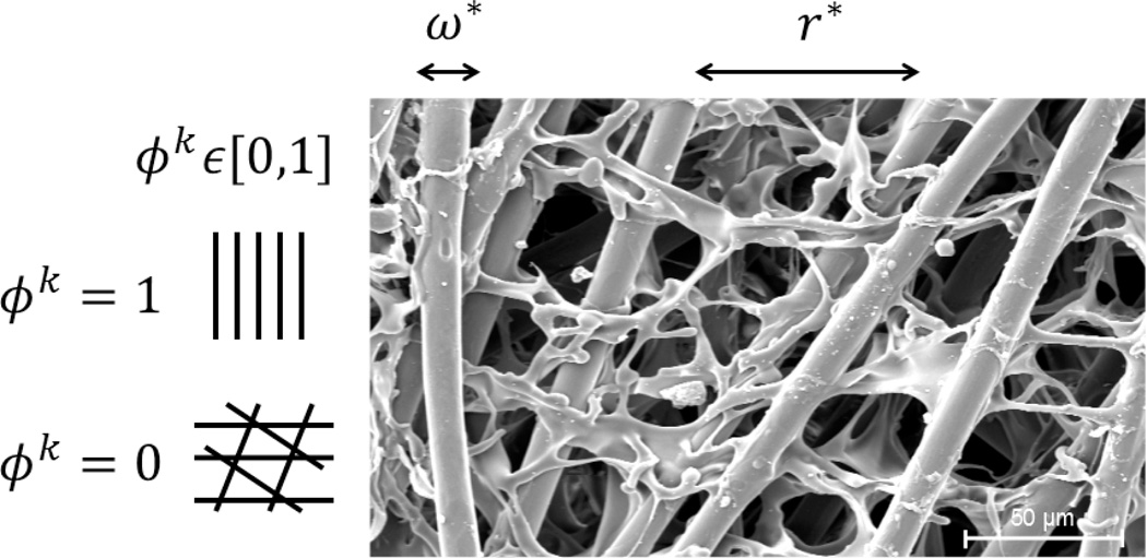
Figure 1.
Scanning electron microscopy image of a PGA-P(CL/LA) scaffold showing the primary parameters considered computationally: normalized fiber diameter ω* and normalized pore size r*. Note that scaffold alignment should also be considered in the future, where ϕp,k = 0 indicates a highly aligned scaffold and ϕp,k = 1 a scaffold with randomly organized fibers. SEM image courtesy of Kevin Rocco.

