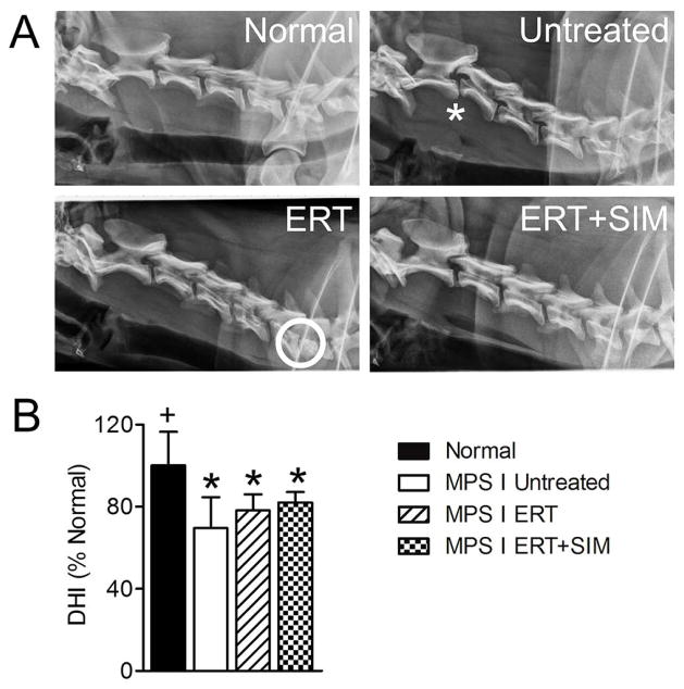
Figure 2. Lateral plain radiographs were obtained for all animals prior to euthanasia at one year-of-age, and analyzed to determine disc height index (DHI).
A. Representative lateral radiographs of the cervical spine for each study group. Asterisk = example of vertebral subluxation and circle = example of collapsed disc space. B. Percent change in DHI calculated from lateral plain radiographs (mean of all levels C2–C7 for each animal). *p<0.05 vs normal; +p<0.05 vs untreated MPS I.

