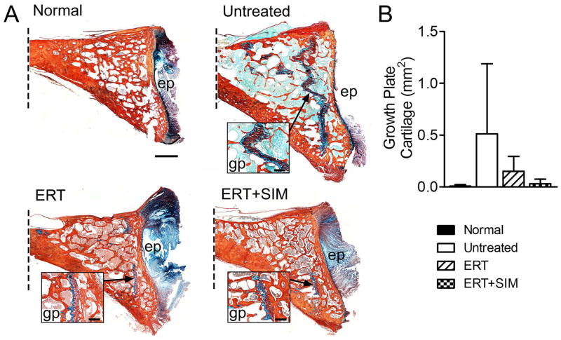
Figure 5. C2 vertebral bodies were processed for histological assessment of remnant growth plate cartilage.
A. Representative histology illustrating remnant growth plate cartilage (insets) in the caudal epiphyses of C2 vertebral bodies (Alcian blue (GAG)/picrosirius red (collagen) stained mid-sagittal sections, oriented with dorsal at the top; dotted line indicates where the image is truncated in the cranial direction). Scale bar = 2mm (main images) and 1mm (insets); ep = vertebral end plate, gp = growth plate. B. Quantification of remnant growth plate cartilage area (mm2) in each study group.

