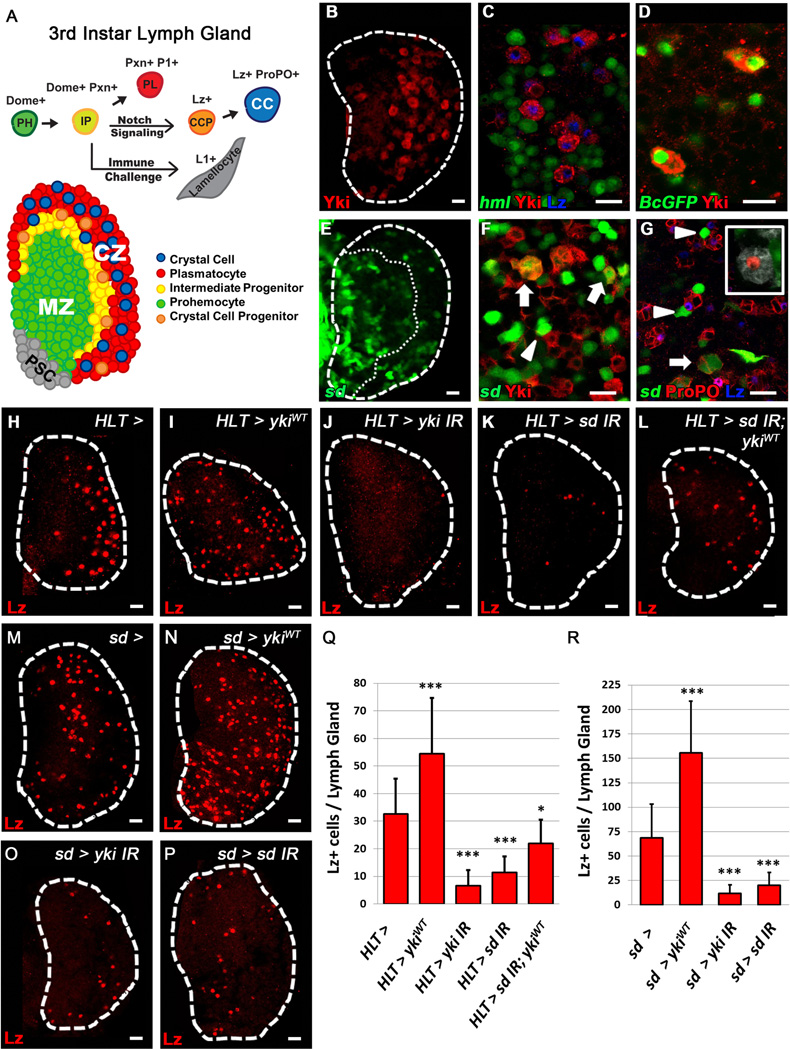
Figure 1. Scalloped and Yorkie are required for proper crystal cell differentiation.
Crystal cell progenitors (CCP) are labeled with Lz (H–P, red). (A) Schematic of the 3rd Instar lymph gland and hemocyte differentiation. PSC in grey, prohemocytes (PH, green) of the MZ, intermediate progenitors (IP, yellow), plasmatocytes (PL, red) and crystal cells (CC, blue) in the CZ. (B) Yki (red) is expressed in scattered cells of the CZ in a 3rd instar lymph gland. (C) Yki (red) is observed in CCPs (Lz, blue) amongst differentiating hemocytes (hml, green) of the CZ. (D) Yki (red) is present in mature CCs labeled with Black cells-GFP (green). (E) sd (sd-gal4 > UAS-2xEGFP, green) is expressed in clusters of cells scattered throughout the lymph gland. CZ is demarcated by a dotted line. (F–G) sd (green) is present in a subset of Yki+ cells (F, arrows) and mature CCs (G, arrows) and is also seen adjacent to Yki+ cells (F, arrowhead) and CCs (G, arrowheads). (G, inset) Lineage traced (sd-gal4, UAS-GFP > UAS-FLP, A5C-FRT-STOP-FRT-LacZ) (red) mature CCs (ProPO, white) do not express sd (green). H–L For each panel, its corresponding pattern of HLT> GFP expression is demonstrated in Fig. S1F–J (H) WT lymph gland (I) Widespread over-expression of ykiWT in the lymph gland increases CCP numbers while (J) depletion of yki or (K) sd blocks CC formation. (L) sd knock-down blocks the increase of CCPs observed upon over-expression of ykiWT. (M) WT lymph gland. (N) Over-expression of ykiWT in sd expressing cells (sd-gal4 >) increases CCP numbers, while (O) depletion of yki or (P) sd strongly inhibits CC differentiation. (Q–R) Quantification of H–P (n=10). * p value <.05, *** p value < .001. Scale bar 10 μm. See also Fig. S1.

