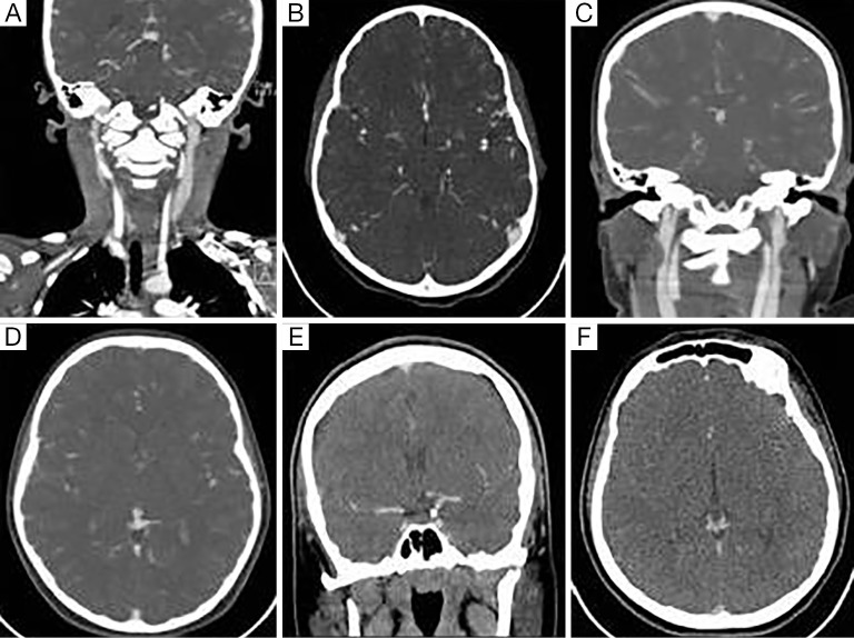
Figure 2.
CTA images of different quality: coronal and axial image of a good quality study (A,B)—note the contrast between the common carotid artery and jugular vein and well delineated cortical arteries. Poor study due to excessive venous contrast (C,D). Poor study with lack of peripheral arterial opacification (E,F). Windowing adjusted for print. CTA, CT angiography.

