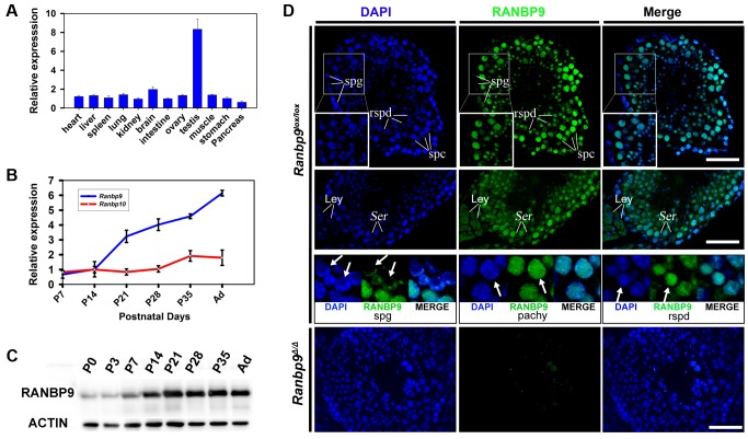
Figure 1. Expression profiles of Ranbp9 during testicular development and spermatogenesis in mice.
(A) qPCR analyses of Ranbp9 mRNA levels in multiple organs in mice. Data are presented as mean ± SEM, n = 3. (B) Expression of Ranbp9 and Ranbp10 during postnatal testicular development. Levels of Ranbp9 and Ranbp10 mRNAs in developing testes at postnatal day 7 (P7), P14, P21, P28, P35, and in adult (Ad) were analyzed using qPCR. Data are presented as mean ± SEM, n = 3. (C) Expression of RANBP9 protein during postnatal testicular development. Levels of RANBP9 in the testes from newborn (P0), postnatal day 3 (P3), P7, P14, P21, P28, and P35 and adult male mice were determined using western blot analyses. ACTIN was used as a loading control. (D) Immunofluorescent detection of RANBP9 in homozygous Ranbp9 flox (Ranbp9lox/lox) and Ranbp9 global knockout (Ranbp9Δ/Δ) testes. In Ranbp9lox/lox testes, RANBP9 immunoreactivity was mostly detected in the nucleus of spermatocytes (spc) and spermatids (spd). Insets show the digitally magnified view of the framed area. RANBP9 was also detected in the nucleolus of Sertoli cells (Ser), and in both the cytoplasm and the nucleus in interstitial Leydig cells (Ley) (Middle panels). While the nucleus was partially RANBP9-positive in a subpopulation of spermatogonia (spg), RANBP9 staining covered the entire nucleus in both pachytene spermatocytes (pachy) and round spermatids (rspd) (Lower panels). In Ranbp9Δ/Δ testes, RANBP9 staining was completely absent. Scale bar = 50 µm.

