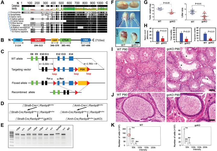
Figure 2. Conditional inactivation of Ranbp9 reveals that male germ cell Ranbp9 is required for normal spermatogenesis and male fertility.
(A) A high degree of conservation of RANBP9 in amino acid sequences, especially at the C-terminus, among ten eukaryotic species. (B) Schematic illustration of the five conserved domains in murine RANBP9, including a proline-rich domain (PRD) at the N-terminus, SPRY, LiSH and CTLH domains in the middle, and a CRA domain at the C-terminus. (C) Schematic representation of the targeting strategy for generating a floxed Ranbp9 allele (Ranbp9lox) through homologous recombination in the murine embryonic stem cells. Exons 12∼14 encode the CRA domain and will be deleted after Cre-mediated recombination. E stands for Exon. Positions of the forward (For) and reverse (Rev) primers used for genotyping are shown. (D) Breeding schemes used for generating germ cell-specific (gcKO) and Sertoli cell-specific (scKO) Ranbp9 conditional knockout mice. (E) Representative PCR genotyping results showing that the floxed (lox) and the WT (+) alleles can be detected as a larger (653 bp) and a shorter (605 bp) bands, respectively. (F) Gross morphology of the testis and the epididymis from WT and gcKO mice at the age of 12 weeks. Scale bar = 1 mm. (G) Testis weight and litter size of 12-week-old gcKO and WT male mice. The gcKO males display significantly reduced testis weight (∼65% of WT) and smaller litter size (∼half of WT) (p<0.05, n = 13). (H) Sperm counts, and total and progressive sperm motility of 12-week-old gcKO and WT male mice, as determined by CASA. Adult gcKO male mice exhibit significantly reduced sperm concentration, total and progressive motility, as compared to age-matched WT mice. Data are presented as mean ± SEM, n = 3. (I) Testicular histology of WT and gcKO mice at postnatal day 90 (P90). Large vacuoles (marked with *) indicative of active depletion of spermatocytes and/or spermatids through sloughing are often seen in seminiferous tubules of the gcKO testes. Scale bar = 50 µm. (J) Epididymal histology of WT and gcKO mice at postnatal day 90 (P90). The WT cauda epididymis is filled with fully developed spermatozoa, whereas the gcKO cauda epididymis contains numerous degenerating/degenerated spermatids or spermatocytes. Scale bar = 60 µm. (K) Flow cytometry-based cell counting analyses showing the altered proportions of three major germ cell types, including haploid spermatids (1n), spermatogonia and somatic cells (2n), and spermatocytes (4n) in WT and gcKO testes. Red circle denotes the fraction representing elongated spermatids and spermatozoa, which is mostly absent in the gcKO testes.

