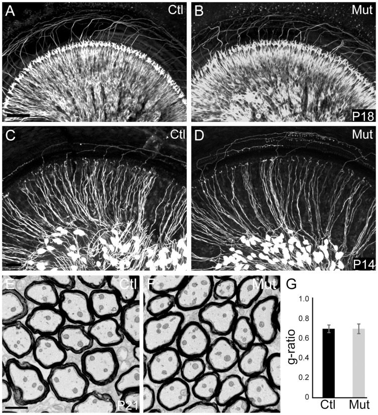
Figure 1. Peripheral SGN connectivity is normal in Npr2 mutant mice.
(A,B) Cochlear innervation was visualized by neurofilament immunostaining of P18 whole cochleae. Visual inspection revealed no obvious difference in the peripheral pattern of connections between wild-type control (Ctl, n = 2) (A) and Npr2 mutant (Mut, n = 2) (B) animals. (C,D) SGN projections in the P14 cochlea were labeled by crossing Neurog1-CreERT2 to the AI14:tdTomato reporter strain, which enables visualization of a subset of SGNs along the length of the cochlea. Individual SGNs of control animals (Ctl, n = 3 heterozygotes) (C) show neatly organized projections within the cochlea. Individual SGN processes of Npr2 mutant (Mut, n = 5) cochleae (D) showed no qualitative differences compared to controls. Note that the degree of labeling can vary slightly independent of genotype, due to fluctuations in Cre activity. (E) Electron micrograph of a transverse section of myelinated SGN axons in the eighth nerve in a control P21 animal. (F) Similar electron micrograph of the eighth nerve of an Npr2 mutant at P21 shows normal axonal diameters and normal myelination. (G) The g-ratio of Npr2 mutants did not differ from controls (P = 0.87, Student's t-test) and was near optimal. Scale bars in A, 50 µm; C, 2 µm.

