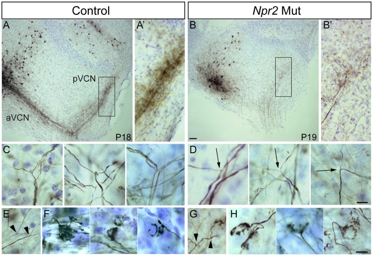
Figure 3. Npr2 mutant SGN axons do not bifurcate properly but can form interstitial branches and morphologically normal synapses.
(A) Injection of biocytin into the aVCN (left) in a parasagittal slice of the cochlear nuclei labeled fibers not only in the nerve root but also in the pVCN (right) in control (Ctl) animals at P18. (A′) A close up view of boxed region in A showing labeled descending branches of SGNs. (B) In an Npr2 mutant (Mut) at P19, biocytin injection into the aVCN also labeled fibers in the nerve root and in the pVCN, showing that at least some fibers branched at the nerve root. (B′) A close up view of the boxed region in B shows labeled SGN fibers and terminals in the pVCN. (C,D) Magnified views of bifurcation zone in control (C) and mutant (D) animals. (C) Examples of stereotyped bifurcations in control animals. (D) In mutants, rare fibers that do branch in the appropriate region do so at irregular angles and with one thinner and one thicker branch (arrows). (E–H) SGN axons can still form interstitial branches and elaborate morphologically normal synaptic endings in Npr2 mutants. Interstitial SGN axon branches, which were distinguished from bifurcations according to morphological criteria, are present in both control (E, arrowheads) and mutant (G, arrowheads) animals. Similarly, endbulbs of Held, which are one terminal whereby SGNs contact bushy cells, show the same types of branching patterns in the control (F) and Npr2 mutants (H). Scale bars in B, 50 µm. Scale bars in D and H, 10 µm. Dorsal is up and rostral is to the left in all panels.

