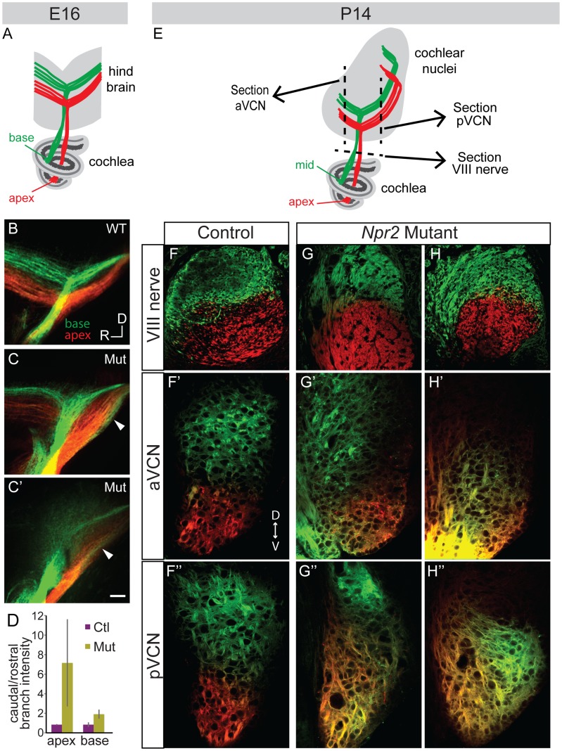
Figure 4. Tonotopy of SGN axons in the cochlear nuclei is blurred in Npr2 mutants.
(A) Schematic diagram illustrating tonotopic dye labeling at E16. Labeling of SGNs in the cochlea with red (apex) and green (base) lipophilic dyes allowed their relative positions to be traced to the hindbrain. (B) In a wild-type (WT) E16 embryo, fibers from the base and apex bifurcated in the nerve root in separate bundles. (C–C′) Similar labeling in two different Npr2 mutants (Mut) shows that fibers from the base and apex were incompletely segregated in the hindbrain. The gross tonotopy was preserved, but some overlap in dye labeling was seen between axons from basal and apical SGNs (arrowheads). Scale bar, 50 µm. (D) The intensity of labeling of SGN axons revealed a caudal bias in Npr2 mutants, with more axons projecting towards the developing pVCN than the aVCN (P<0.05, Student's t-test). (E) Schematic diagram illustrating tonotopic dye labeling at P14. The planes of sections illustrated in F–H are indicated by dotted lines. (F–F″) In a control animal, axons arising from the middle and from the apex of the cochlea were segregated in the auditory nerve and in the aVCN and pVCN, as assessed qualitatively using confocal imaging. (G–G″, H–H″) Two examples of Npr2 mutants. Axons arising from the middle and apical turns of the cochlea were properly segregated in the nerve, indicating that the dyes labeled physically distinct populations of neurons as in controls (G,H). However, axons from these neurons were intermingled in the aVCN (G′, H′) and pVCN (G″, H″) of the same animals.

